Activity of our biophoton research has been introduced in the "NewScietist" Journal. This is the copy of the article published on 23 Feb. 2002 issue.
|

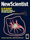
Can faint flashes of light tell us how cells are feeling? And if we learn their language, could we ever take control, asks Bennett Daviss MASAKI KOBAYASHI is looking for the inner light in all of us. A physicist at the Tohoku Institute of Technology in Sendai, Kobayashi is one of a small number of researchers around the globe who spend hours locked in darkened labs in the hope of glimpsing the faint glow that comes from all living tissue. This glow is so elusive that we can only see it with the aid of the most sensitive detectors. But we're not talking about psychic auras here. These faint throbs of light are probably little more than a by-product of the cell's own metabolism-the pint-sized equivalent of a spluttering car exhaust. Yet researchers hope that one day differences in the light from healthy and cancerous cells will give doctors a new non-invasive tool for spotting disease. Others go further. They think that cells may coordinate their activities via patterns of photons. A few even dare to suggest that these photons may actually mediate consciousness itself. Living creatures can undoubtedly emit light: around 80 per cent of marine creatures, for example, fireflies and quite a few fungi and centipedes use bioluminescence as their calling card. Generating this light usually involves a chemical reaction between ATP-the cell's energy store-oxygen and a molecule called luciferin. Luciferin converts the chemical energy locked up in ATP into photons of light (New Scientist, 22 July 2000, p 34). In most cases these cells produce a weak flash that is just visible to the human eye. But the kind of light that Kobayashi is interested in is far weaker-it is typically several million times as faint as the light from a firefly. In fact, the trickle of "biophotons" is so weak that researchers are only now beginning to agree where it comes from. The main source is free radicals: atoms or molecules with an unbound electron that are desperate to pair up with electrons from other molecules. Free radicals are often an unwelcome by-product of the reactions that take place at the inner membrane of mitochondria-the power houses of the cell that use oxygen to make the cell's fuel ATP. Free radicals are seriously bad news. When they bump into other molecules in the cell such as proteins, lipids or sugars, they destroy them by slicing them up into small chunks. Most biological reactions take place in several small steps, each one designed to use energy efficiently. But these free radical reactions are so energetic that they tend to occur in one huge step. This means not all the energy is used up in the reaction. A little is absorbed by an electron on the molecule that's under attack. This electron becomes unstable and sheds its extra energy as a photon of light. Since enzymes and anti-oxidants usually mop up reactive oxygen molecules and free radicals before they can damage the cell, a healthy cell tends to release very few photons, maybe only tens per minute. Not easy to collect, even in a pitch-black lab. This is one of the reasons the phenomenon has been so difficult to study. In the 1970s, biochemists first considered biophotons as a way of studying reactive oxygen molecules. "However biophoton emission is so weak and the mechanisms of production are so complex, most biochemists were put off," Kobayashi says. For example, veteran biophysicist Britton Chance at the University of Pennsylvania, Philadelphia, showed that light was coming from free radicals created in isolated mitochondria. "But detailed studies failed to detect a signal in dog's brain," he says. Things got a little easier in the 1980s when manufacturers such as Hamamatsu, a Japanese company specialising in light detectors called photomultipliers, developed new highly sensitive instruments designed to record weak light signals. Keen to exploit this opportunity, and to seed a new bio-optics industry, the Japanese government funded a five-year, multibillion-yen research programme into biophotons in 1986. Humio Inaba, an engineer at the Research Institute of Electrical Communication at Tohoku University headed the project. Dozens of researchers across Japan, including Kobayashi, found these emissions coming out of everything from plant seeds to fruit flies. Inaba also discovered that injured or stressed cells release far more photons than their healthy counterparts. In particular, adzuki and soybean seedlings damaged with cross-shaped cuts emit high levels of photons at the site of the injury. Other teams have spotted increased levels of biophotons where cells are damaged. Ken Muldrew, a biophysicist at the University of Calgary in Alberta, Canada, tore tree leaves apart near his sensitive measuring equipment: "We got an enormous peak of tens of thousands of photons, a burst of light," says Muldrew. "A leaf screams when you tear it, but you see the scream instead of hear it." It isn't just plant cells. At the Institute of Physics at the University of Catania in Italy, isolated mammalian tumour cells ejected photons at rates as high as 1400 per square centimetre per minute-healthy tissues average rates of less than 40. In a study on bladder cancer, Kobayashi's team found that the light intensity of tumour cells is 4 times as high as the surrounding healthy tissue. Clearly when cells are stressed or damaged, they pump out free radicals-and this produces light. But can doctors use these distress flares as warnings of disease or illness? Almost certainly, says Reiner Vogel, a biophysicist at the University of Freiburg in Germany. "The emission may give a very sensitive indication of the conditions within a cell and on the functioning of the cellular defence mechanism," he says. Philip Coleridge Smith, a surgeon at University College Medical School in London, agrees. You could perhaps use biophotons to assess inflammation in tissues, he suggests, which might warn of leg ulcers, for example. To make a diagnosis what we need now is a sensitive detector and analysis system, preferably non-invasive, that will measure an emission and even identify its origins-perhaps from the spectrum or statistical properties of the photons, Kobayashi says. He's trying to find out if you can use photons to spot disease in people, rather than in cells in a lab, and is developing ways to convert patterns of photon emissions into images of the body that resemble X-rays or CAT scans. Turn to the light Yet some believe that biophotons are far more than just distress signals. In the early 1990s, Guenter Albrecht-Buehler, a biophysicist at Northwestern University Medical School in Chicago, discovered that some cells can detect and respond to light from others. He shone infrared light onto a mixture of cell-sized latex beads and mouse fibroblast cells. Many of the cells began to stretch out their arm-like pseudopodia for light scattered towards them by the beads, and soon these cells were heading directly for the beads. Some even turned 180° to reach them. (With little power and a wavelength of around 850 nanometres, the light created virtually no heat, so the cells weren't simply moving towards warmth, argues Albrecht-Buehler.) And since some cells reached out to two different light sources of equal intensities at the same time, it seems that they could "see" each source distinctly, he suggests. In other experiments, Albrecht-Buehler spread hamster cells on both sides of a sheet of glass. As the cells grew, he found that those on one side shifted around until they lay at angles of more than 45° to those on the other side of the glass. But when he added a filter layer to the glass that blocked infrared light transmission from one side to the other, the cells grew in random directions (New Scientist, 7 November 1992, p 14). Tissues favour a criss-cross arrangement of cells because it gives them extra strength, so perhaps the cells on the glass were using light to signal their orientation. If so, they must have some kind of eye. Albrecht-Buehler thinks the cell's centrioles fit the bill. These cylindrical structures have slanted "blades" which he believes act as simple blinds. By only allowing light into the centriole from certain angles, the blinds enable simple photoreceptors inside the centrioles such as haem molecules to tell which direction photons are coming from. And microtubules-hollow filaments that thread through cells-could act as optical fibres, he believes, feeding light towards the centrioles from the cell's wall. But why should cells want to detect light? The most obvious answer is that they are talking to each other, says Albrecht-Buehler. Cells in embryos might signal with photons so that they know how and where they fit into the developing body. And now he wants to learn their language. He envisages doctors telling cells what they want them to do in words they understand. You might tell cancer cells to stop growing or encourage cells near wounds to start again. "We may learn to compose our own messages in the language of cells to compel them to carry out specialised tasks that they've never performed." Albrecht-Beuhler isn't the first to make this controversial claim. In the 1980s Fritz-Albert Popp, then a lecturer at the University of Marburg in Germany, became interested in the optical behaviour of cells. In a series of experiments Popp found that two cells separated by an opaque barrier release biophotons in uncoordinated patterns. Remove the barrier and the cells soon begin releasing photons in synchrony. The cells, Popp concluded, were communicating by light. Cyril Frank, professor of surgery at the University of Calgary's medical school believes Popp could be right. A photon could trigger events in the receiving cell, making it change its rate of division or express different proteins, he suggests. "In our experiments, we're trying to find out if these sorts of triggers can do things like that." Although not ready to disclose any data, he says they're "getting some encouraging results". Keep it simple But Muldrew feels biophotons can only communicate simple messages. "What biophotons communicate is the fact that certain oxidative reactions are going on." It's hard to know whether glowing cells will ever shed light on disease, help scientists to work out the language of cells or even whether biophotons play a role in consciousness (see "Inner Light"). Cells are simply far too sensitive to factors that alter the rate at which photons are emitted. "The problem is reproducibility of results, even for relatively simple systems like cell cultures," says Barbara Chwirot, head of the Laboratory of Molecular Biology of Cancer at Nicolas Copernicus University in Torun, Poland. Light emission may depend on free radicals, but it is also affected by enzyme activity and the supply of protective antioxidants such as vitamin E or carotenoids. "Direct diagnosis of disease will remain difficult without some technical or medical breakthrough," says Kobayashi. For now the focus is on more prosaic pursuits. Popp, who now heads the International Institute of Biophysics in Neuss, Germany, an association of scientists interested in biophoton research, runs a company called Biophotonen that offers its expertise in reading photon emissions to gauge the freshness and purity of food. One of its first projects is with German brewer Bitburger, the idea being to spot the glow from harmful bacteria before they can get into its beer. A Chinese research group in Beijing is also perfecting a photon-based test for the presence of microbes that could be used in the food industry. But one day soon, bugs may not be the only things under the lens. Kobayashi hopes that a new generation of detectors, such as avalanche photodiodes that boost the chances of detecting individual photons from around 20 per cent up to 80 per cent or more, could provide the breakthrough he needs to develop a scanner to diagnose disease. It might take 10 or 20 years, Kobayashi says, but at last he thinks he can see light at the end of the tunnel. Inner lightSome maverick theorists suggest that the notion of a common cellular language puts biophotons at the centre not only of biological communication, but also of consciousness. Scott Hagan, a theoretical physicist at the British Columbia Institute of Technology in Burnaby, has been pondering the enigmas of awareness since he shared an office in grad school with a neurophysiologist. "In every other process, every atom or molecule does its own thing," he says. However, we cannot think unless cells in the brain function simultaneously, as parts of a whole. There's no way that classical physics can explain this, he says. "But in quantum physics, there are systems that know they're wholes. These are called quantum coherent states." These are states in which the wave functions of atoms or molecules blend to form one single unit (New Scientist, 24 March 2001, p 42). And the search for quantum coherent states in the brain leads inside individual neurons, says Hagan, to their skeleton-like framework of microtubules. These thin tubes are thought to move energy about the cell, help build junctions between neurons and maintain memory. And anaesthetics work by binding to them, says Hagan. "Because anaesthetics make consciousness evaporate, their site of action is important in determining the mechanisms responsible for consciousness," Hagan says. In a highly speculative theory, Hagan and Stuart Hameroff, associate director of the Center for Consciousness Studies at the University of Arizona, suggest that quantum coherence in the protein subunits of microtubules may give rise to consciousness (New Scientist, 20 August 1994, p 35). And, says Hameroff, biophotons could somehow control this process.
Bennett Daviss is a science writer in New Hampshire |
|||
| (c) Copyright New Scientist, RBI Limited 2001 |